Advances in catheter technologies have enabled new diagnostic and therapeutic procedures that continue to transform healthcare. These advances are led by innovations in catheter design that push the boundaries of materials science and introduce new technologies and capabilities.
The standard makeup of a catheter is the starting point for developing any new catheter device. In this blog, we’ll look at the makeup of catheters used in interventional radiology procedures as well as typical performance characteristics.
Catheter Applications in Interventional Radiology Procedures
- Angiography – where catheters deliver a contrast agent into blood vessels that can then be visualised using a variety of imaging techniques to detect, for example, blockages, aneurysms, or malformations in blood vessels.
- Angioplasty and stenting – where catheters deliver a small balloon or stent to a narrowed or blocked blood vessel. The balloon is then inflated to open the vessel, and the stent is left in place to keep it open.
- Embolization – where catheters are used to deliver agents that block a blood vessel. This can be used to cut off blood supply to a tumour, stop bleeding, or treat aneurysms.
- Thrombolysis – where catheters are used to deliver clot-dissolving medications directly to the site of a clot.
- Chemoembolization and radioembolization – where catheters are used to deliver chemotherapy drugs or radioactive materials directly to a tumour, often combined with an embolizing agent to restrict blood flow and keep the therapeutic agent in the tumour for longer.
- Drainage procedures – where catheters are used to drain fluids, including for conditions such as abscesses or pleural effusions.
- Central venous access – where catheters are used to provide long-term access to a patient’s bloodstream for the administration of medications, nutrition, or blood products.
- Ablation procedures – where catheters are used to deliver energy (like radiofrequency, microwave, or cryoablation) to ablate, or destroy, abnormal tissues. Ablation procedures are often used to treat certain types of cancer or cardiac arrhythmias.
Interventional Radiology Catheters: Performance Characteristics
Materials
Catheters must be made using biocompatible materials that won’t cause reactions when inserted into the body. Medical-grade plastic polymers, such as polyurethane, Nylon, silicone, and polyethylene, are commonly used materials. The materials selected for a catheter have an impact on the performance characteristics of the device, including flexibility, durability, and resistance to various chemicals and body fluids.
Size
Catheter size is described in terms of length and diameter. The length of a catheter is usually based on the part of the body it is to be inserted into, while the diameter (often referred to in terms of its French size) is selected based on the specific procedure being performed. Wall thickness is another crucial consideration, as ultra-thin walls minimise the overall diameter of a catheter while still allowing the maximum lumen size.
Radiopacity
Catheters used in interventional radiology are often radiopaque to make them visible under imaging modalities such as X-rays or CT scans. This helps interventional radiologists guide the catheter to the correct location in the body. The radiopacity typically is achieved by using fillers in the catheter walls or using marker bands along the length of the catheter.
Torqueability and Trackability
Torqueability and trackability refer to the ability of the catheter to navigate through the vascular system. Torque is the ability to rotate the catheter’s distil tip by turning it from the proximal end, while trackability refers to the ease with which a catheter can follow a guide wire.
Lubricity
Catheters are often made using low friction materials or have hydrophilic coating added to make them more slippery. The reduced friction aids insertion and helps prevent damage to vessel walls while the device moves through the body.
Flexibility and kink resistance
Catheters need to be flexible enough to navigate the complex structure of the body’s vascular system, but also rigid enough to resist kinking or collapsing. Composite catheters can have multiple durometer materials and reinforcement along the shaft length of the device to give it the required properties.
Multi-lumen design
Some catheters have multiple lumens, or channels, allowing them to perform multiple functions at once, such as delivering drugs, injecting contrast, or measuring pressures.
The Makeup of Composite/layered Catheters Used in Interventional Radiology Procedures
Innermost Layer: Core or Liner
The innermost layer of a catheter is usually a thin, flexible core or liner made of materials such as polytetrafluoroethylene (PTFE) or thermoplastics. This layer provides a smooth pathway for medical instruments or wires to navigate through the catheter during the procedure.
Reinforcement Layer
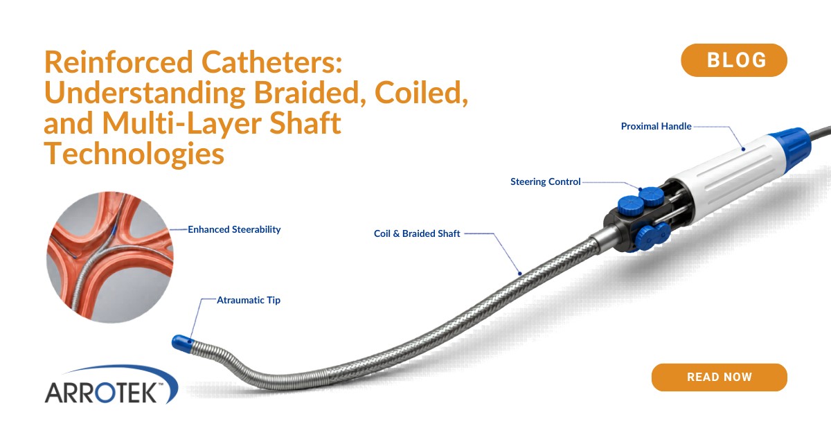
The reinforcement layer is responsible for providing structural support as well as helping to prevent the catheter from kinking or collapsing. The reinforcement layer is often made of braided and/or coiled stainless steel or nitinol wires. These wires add flexibility and strength to the catheter, allowing it to navigate through tortuous anatomies. Variation in coil and braid density along the shaft length of the device can be used to give it the required properties such as kink resistance, torqueability and tensile strength.
Intermediate Layers
In some catheters, there may be additional intermediate layers between the reinforcement layer and the outermost layer. These layers can serve various purposes, such as enhancing flexibility, adding radiopacity, improving lamination, or improving torque transmission for better manoeuvrability.
Outermost Layer: Polymer Jacket
The outermost layer, also known as the polymer jacket, encapsulates the reinforcement and intermediate layers. It is typically made of a biocompatible thermoplastic such as polyurethane, nylon or derivatives. This layer provides a smooth surface, protecting the inner layers and facilitating the passage of the catheter through blood vessels or other body structures. This layer can include fillers/additives to increase radiopacity and reduce friction.
Marker Bands or Radiopaque Markers
In some catheters, small metal bands or markers made of materials like platinum, stainless steel or gold are placed along the length of the catheter. These markers are highly radiopaque to make them visible under X-ray fluoroscopy. Marker bands and radiopaque markers help physicians accurately track the position and movement of the catheter within the body during the procedure.
Distal Tip
The distal tip of a catheter is one of the most specialised parts. It can be designed in various shapes, such as a straight or angled. Distil tip shapes can also be designed for specific applications. The distal tip is often flexible and atraumatic, minimising the risk of tissue damage during navigation through delicate vessels or anatomical structures. Some catheters also have inflatable balloons near the tip that can be used to anchor the catheter in place or apply treatments directly to certain areas.
Catheter Design Support at Arrotek
At Arrotek, we offer design expertise for catheters used in interventional radiology procedures. This includes everything from supporting part of your project to managing the full design process. To discuss your requirements, please get in touch today.